Home

Rohöl am wenigsten Jugend mri swan sequence Bibel Lager Optimal

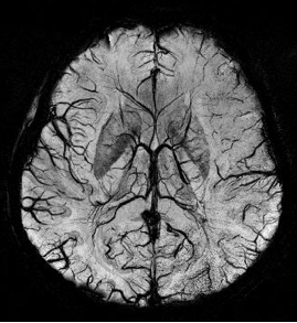
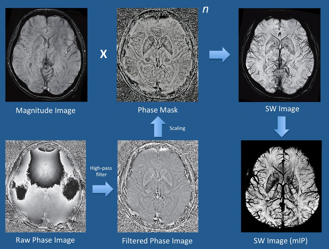
Susceptibility phase - Questions and Answers ​in MRI

Susceptibility weighted imaging - Wikipedia

Susceptibility weighted imaging | Radiology Reference Article |  Radiopaedia.org
Susceptibility weighted imaging | Radiology Reference Article | Radiopaedia.org

PDF] SWAN sequence in comparison to T2 for STN visualisation in DBS surgery  | Semantic Scholar
PDF] SWAN sequence in comparison to T2 for STN visualisation in DBS surgery | Semantic Scholar

Magnetic Resonance Imaging (MRI) | Radiology Key

Frontiers | Diffusion- and Susceptibility Weighted Imaging Mismatch  Correlates With Collateral Circulation and Prognosis After Middle Cerebral  Artery M1-Segment Occlusion
Frontiers | Diffusion- and Susceptibility Weighted Imaging Mismatch Correlates With Collateral Circulation and Prognosis After Middle Cerebral Artery M1-Segment Occlusion

SWAN-Venule: An Optimized MRI Technique to Detect the Central Vein Sign in  MS Plaques | American Journal of Neuroradiology
SWAN-Venule: An Optimized MRI Technique to Detect the Central Vein Sign in MS Plaques | American Journal of Neuroradiology

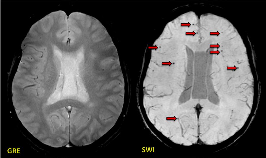
Superiority of SWI over GRE - Questions and Answers ​in MRI

SWAN Brain Imaging by KMH Labs - Issuu

SWI, susceptibiltiy - Questions and Answers ​in MRI

Figure 2 from Diagnosis of intracranial hemorrhagic lesions: comparison  between 3D-SWAN (3D T2*-weighted imaging with multi-echo acquisition) and  2D-T2*-weighted imaging | Semantic Scholar
Figure 2 from Diagnosis of intracranial hemorrhagic lesions: comparison between 3D-SWAN (3D T2*-weighted imaging with multi-echo acquisition) and 2D-T2*-weighted imaging | Semantic Scholar
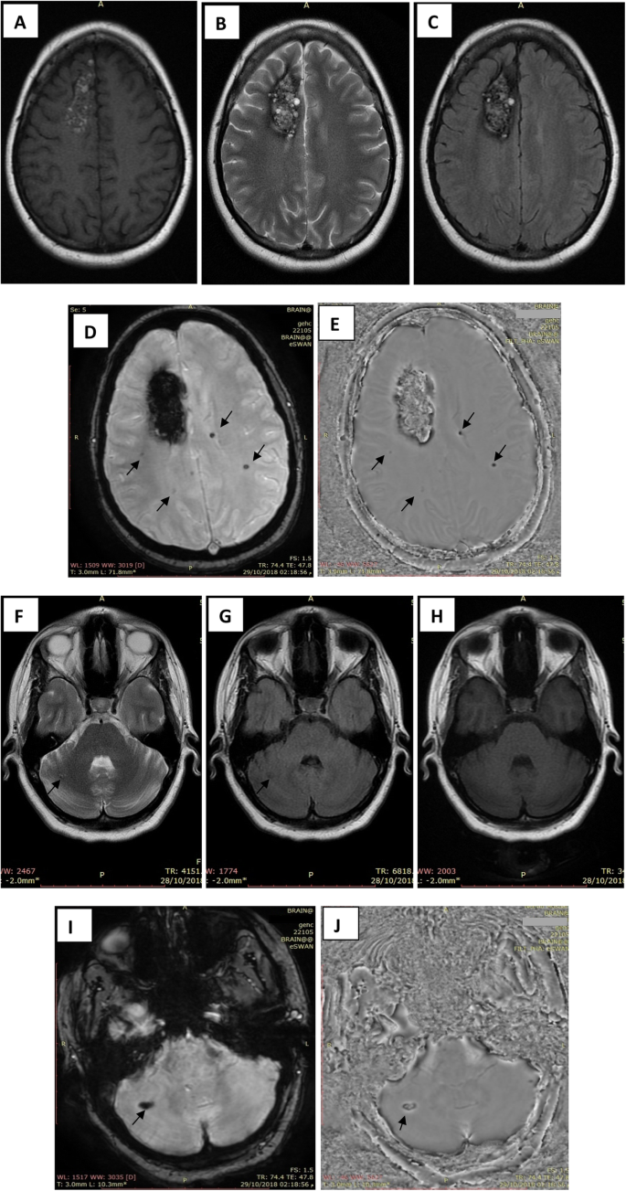
Magnetic resonance susceptibility weighted in evaluation of cerebrovascular  diseases | Egyptian Journal of Radiology and Nuclear Medicine | Full Text
Magnetic resonance susceptibility weighted in evaluation of cerebrovascular diseases | Egyptian Journal of Radiology and Nuclear Medicine | Full Text

Susceptibility-weighted Imaging: Technical Essentials and Clinical  Neurologic Applications | Radiology
Susceptibility-weighted Imaging: Technical Essentials and Clinical Neurologic Applications | Radiology

Susceptibility-weighted Imaging: Technical Essentials and Clinical  Neurologic Applications | Radiology
Susceptibility-weighted Imaging: Technical Essentials and Clinical Neurologic Applications | Radiology

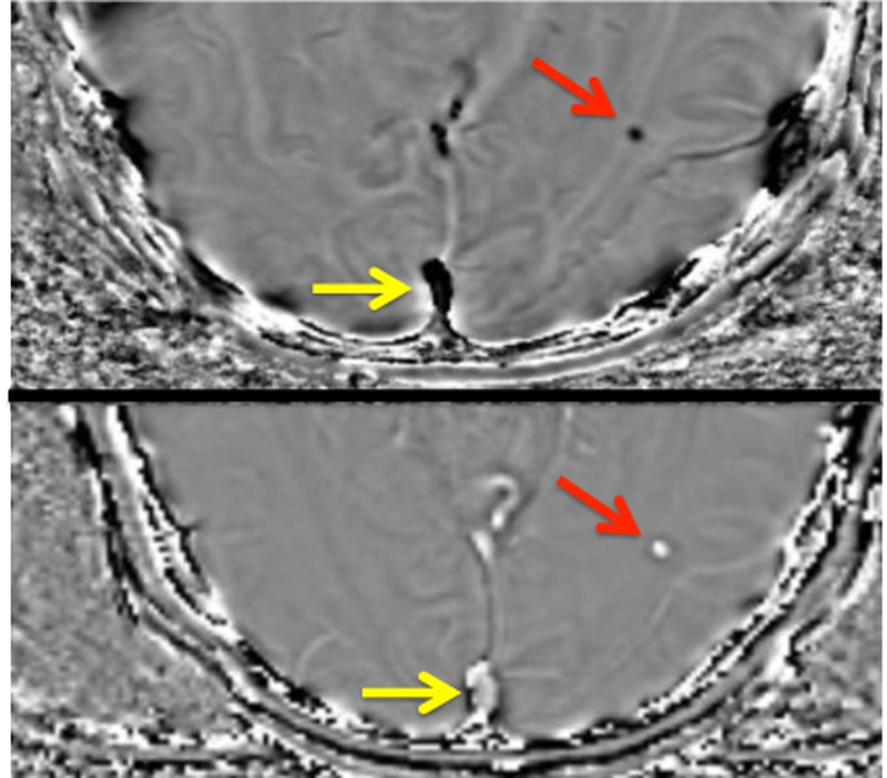
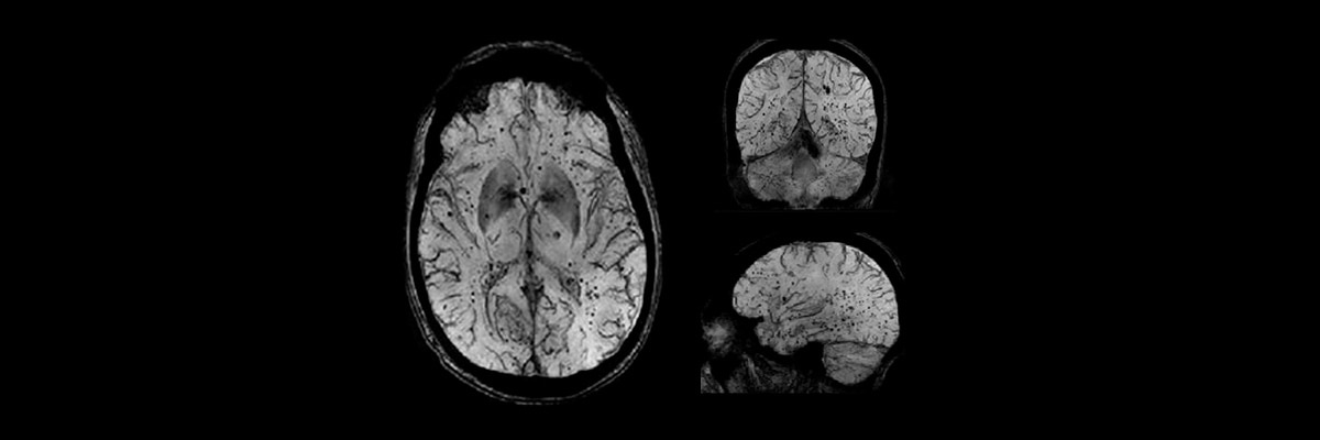
GE Healthcare See brain vasculature in exquisite detail in one quick, easy  scan.
GE Healthcare See brain vasculature in exquisite detail in one quick, easy scan.

MRI Technologist - MR Imaging of the brain in a patient with glioblastoma  multiforme (GBM). High resolution anatomical sequences (volumetric T2,  volumetric FLAIR, volumetric T1), 3D SWAN (for hemorrhage and vasculature  assessment)
MRI Technologist - MR Imaging of the brain in a patient with glioblastoma multiforme (GBM). High resolution anatomical sequences (volumetric T2, volumetric FLAIR, volumetric T1), 3D SWAN (for hemorrhage and vasculature assessment)

Susceptibility-weighted angiography (SWAN) of cerebral veins and arteries  compared to TOF-MRA
Susceptibility-weighted angiography (SWAN) of cerebral veins and arteries compared to TOF-MRA

Comparison between SWAN MRI sequences before and after early... | Download  Scientific Diagram
Comparison between SWAN MRI sequences before and after early... | Download Scientific Diagram

SWAN-targeted axial image of the midbrain in a healthy subject... |  Download Scientific Diagram
SWAN-targeted axial image of the midbrain in a healthy subject... | Download Scientific Diagram

SWAN MR Application | GE HealthCare (United States)