Home

Strand Anbetung behindert mri abdomen sequences Observatorium Grenze Besitz

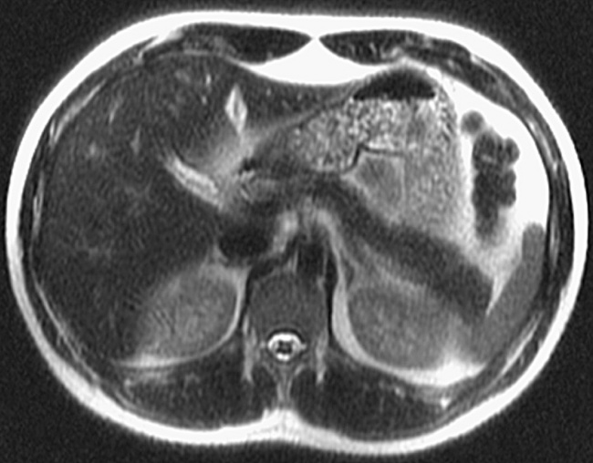
Abdominal MRI (T2-weighted sequence, axial slice), showing a lobulated cyst  located in the body of the pancreas.
Abdominal MRI (T2-weighted sequence, axial slice), showing a lobulated cyst located in the body of the pancreas.

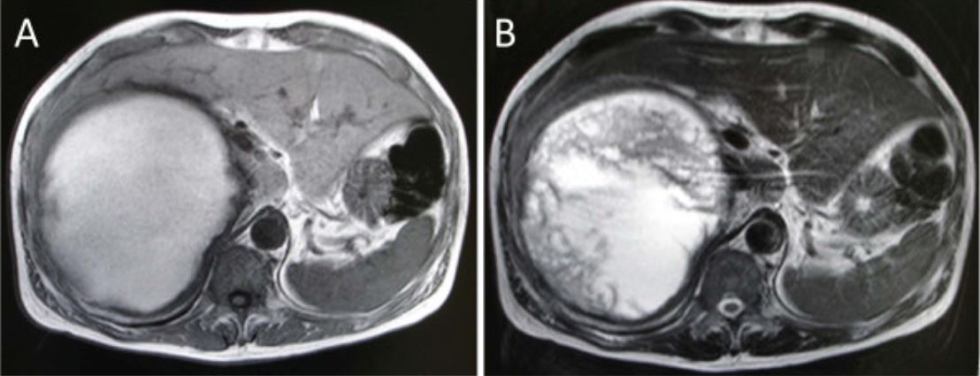
MRI sequences. (A) T1 shows numerous lesions with T1 shortening. (B) T2...  | Download Scientific Diagram
MRI sequences. (A) T1 shows numerous lesions with T1 shortening. (B) T2... | Download Scientific Diagram

DCE performance - Questions and Answers ​in MRI

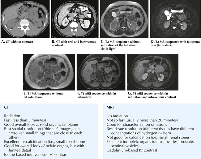
Abdomen | Radiology Key

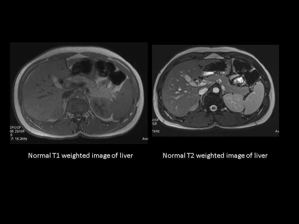
Abdominal MRI T1 (A,B) and T2( C,D) sequences A and C are healthy... |  Download Scientific Diagram
Abdominal MRI T1 (A,B) and T2( C,D) sequences A and C are healthy... | Download Scientific Diagram

This is a basic abdominal MRI protocol when using an extracellular... |  Download Scientific Diagram
This is a basic abdominal MRI protocol when using an extracellular... | Download Scientific Diagram

Proposed MRI protocol: sequences and rationale, with examples of how to...  | Download Scientific Diagram
Proposed MRI protocol: sequences and rationale, with examples of how to... | Download Scientific Diagram

BioMedInformatics | Free Full-Text | Improving Deep Segmentation of  Abdominal Organs MRI by Post-Processing
BioMedInformatics | Free Full-Text | Improving Deep Segmentation of Abdominal Organs MRI by Post-Processing

Abdominal wall mri protocol, planning and indications | endometriosis  anterior abdominal wall mri
Abdominal wall mri protocol, planning and indications | endometriosis anterior abdominal wall mri

Abdominal MRI (T2-weighted sequence with fat saturation, axial slice),  showing a lobulated cyst located in the uncinate process of the pancreas.
Abdominal MRI (T2-weighted sequence with fat saturation, axial slice), showing a lobulated cyst located in the uncinate process of the pancreas.

MR Abdomen, MRI of Pelvis, MRI of Abdomen, MRI Scan

MRI of the Abdomen and Pelvis | Radiology Key

Abdominal MRI (T2-weighted sequence with fat saturation, axial slice),  showing two small cysts, one in the tail and one in the transition between  body and tail of pancreas.
Abdominal MRI (T2-weighted sequence with fat saturation, axial slice), showing two small cysts, one in the tail and one in the transition between body and tail of pancreas.

Abdominal MR Imaging at 3.0 T | RadioGraphics

Abbreviated MRI Protocols for the Abdomen - YouTube

Abdominal MRI T2 fast field echo sequences. (A, C, E) Normal pancreas... |  Download Scientific Diagram
Abdominal MRI T2 fast field echo sequences. (A, C, E) Normal pancreas... | Download Scientific Diagram

MR scanning - Questions and Answers ​in MRI

EPOS™
EPOS™

RAVE-T2/T1 – Feasibility of a new hybrid MR-sequence for free-breathing abdominal  MRI in children and adolescents - European Journal of Radiology
RAVE-T2/T1 – Feasibility of a new hybrid MR-sequence for free-breathing abdominal MRI in children and adolescents - European Journal of Radiology

Magnetic Resonance Imaging (MRI) | Concise Medical Knowledge

Dixon method - Questions and Answers ​in MRI

Abdominal wall mri protocol, planning and indications | endometriosis  anterior abdominal wall mri
Abdominal wall mri protocol, planning and indications | endometriosis anterior abdominal wall mri

Abdominal MRI T2 FFE (fast field echo) sequences show hypointense... |  Download Scientific Diagram
Abdominal MRI T2 FFE (fast field echo) sequences show hypointense... | Download Scientific Diagram

Abdominal wall mri protocol, planning and indications | endometriosis  anterior abdominal wall mri
Abdominal wall mri protocol, planning and indications | endometriosis anterior abdominal wall mri

PDF] Simulation of abdominal MRI sequences in a computational 4 D phantom  for MRI-guided radiotherapy | Semantic Scholar
PDF] Simulation of abdominal MRI sequences in a computational 4 D phantom for MRI-guided radiotherapy | Semantic Scholar

Figure 1—29 from Free-breathing 3D T1-weighted gradient-echo sequence with  radial data sampling in abdominal MRI: preliminary observations. | Semantic  Scholar
Figure 1—29 from Free-breathing 3D T1-weighted gradient-echo sequence with radial data sampling in abdominal MRI: preliminary observations. | Semantic Scholar

Diagram illustrating how to differentiate T1-weighted, T2-weighted, and...  | Download Scientific Diagram
Diagram illustrating how to differentiate T1-weighted, T2-weighted, and... | Download Scientific Diagram

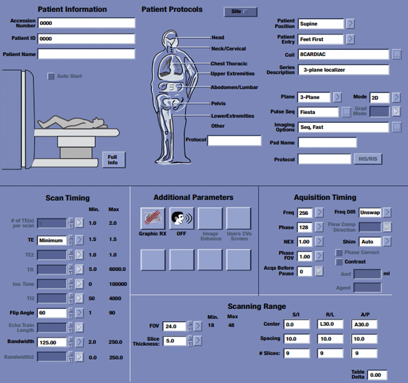
Abdominal MRI images: (A) A coronal FIESTA sequence showing the... |  Download Scientific Diagram
Abdominal MRI images: (A) A coronal FIESTA sequence showing the... | Download Scientific Diagram